Squamous Cell Carcinoma Treatment in Sarnia, Ontario
Squamous cell carcinoma (SCC) is the second most common skin cancer.
At a glance
- OHIP
- OHIP-covered
Usually, when it is medically necessary
No referral needed.
Squamous cell carcinoma (SCC) is the second most common skin cancer. Most are found and treated before they spread, but SCC can grow faster than basal cell carcinoma, and a small share can spread. It is assessed with care and treated promptly.
SCC in situ (Bowen’s disease)
An early form that is still in the top layer of skin
Invasive SCC
Has grown into the deeper skin layers
Often on sun-exposed skin
Face, ears, lips, scalp, hands and lower legs
Confirmed by biopsy
The depth and features are checked before treatment is planned
Treated in clinic when suitable
Some high-risk cases are referred
Quick answers
Is SCC serious?
Most are treated successfully, especially when found early. Some have features that raise the risk of return or spread.
What is SCC in situ?
An early SCC confined to the top layer of skin. It is also called Bowen’s disease.
Do I need a biopsy?
Yes, to confirm the diagnosis and to see how deep the cancer goes
What is the usual treatment?
Surgical excision with a margin. Some in situ and thin lesions can be treated in other ways.
Will it leave a scar?
Yes, every method leaves some mark
Can it come back?
Yes. Follow-up skin checks are advised.
Is it covered by OHIP?
Usually, when it is medically necessary
What are squamous cell carcinoma and SCC in situ?
SCC begins in the squamous cells, the flat cells that make up most of the top layer of skin (the epidermis). Non-melanoma skin cancers are the most common cancers in Canada, and about 20% of them are SCC.
SCC in situ (Bowen’s disease)
Abnormal cells are limited to the epidermis. They cannot spread to lymph nodes or other organs at this stage. If left, it can grow into invasive SCC over time.
Invasive SCC
The cancer has grown into the deeper layer of skin (the dermis) or beyond. It can occasionally spread.
Actinic keratosis (AK)
A rough, scaly sun-damage patch. It is not a cancer, but a few can progress to SCC.
Causes and risk factors
- Years of UV exposure from the sun and tanning beds
- Fair skin and a history of sunburn
- Earlier actinic keratoses or skin cancer
- A weakened immune system, for example after an organ transplant or with some medicines
- Chronic wounds, burn scars or long-standing skin inflammation
- Older age
Anyone can develop skin cancer, whatever their skin colour.
What can squamous cell carcinoma look like?
SCC has several looks. Only an examination can tell it apart from other skin conditions, so this list is a guide to when to book a check, not a way to diagnose.
A red, scaly or crusted patch
Slow-growing and often dry. SCC in situ commonly looks like this.
A firm, pink or red bump
May have a rough or crusted top.
A wart-like growth
May bleed easily.
A sore that does not heal
May crust, ooze or reopen.
A rough patch on the lip
Needs checking, because the lip is a higher-risk place.
A growth that rises quickly over weeks
Should be seen soon.
Book a check if you notice
- A scaly patch that persists or keeps coming back despite moisturizer
- A sore, bump or crust that does not heal in 4 weeks
- A growth that is tender, bleeds or is getting larger
- A change in an old scar, burn or ulcer
SCC is more often found on sun-exposed skin, but it can occur anywhere on the body.
How squamous cell carcinoma is assessed and diagnosed
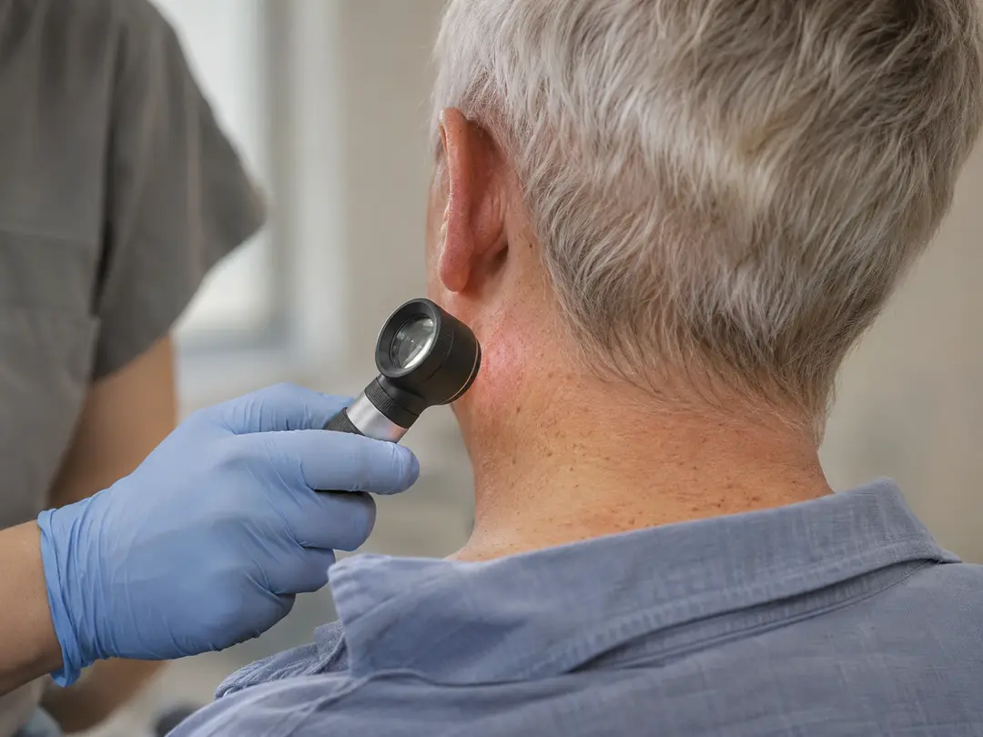
All skin lesions are assessed by a doctor before treatment. The assessment combines clinical experience, dermoscopy and digital imaging. AI-assisted analysis is used where appropriate, alongside the doctor's own judgement.
- 1
History
How long the spot has been there, changes, symptoms, sun exposure, past skin cancer and medicines that affect the immune system
- 2
Full skin examination
Sun-exposed and covered areas are checked. Nearby lymph nodes may be felt if an invasive SCC is suspected.
- 3
Dermoscopy
A lit magnifier shows structures and vessel patterns that help separate SCC, AK and other lesions
- 4
Biopsy
A sample is taken under local anesthetic and sent to a lab. It confirms the diagnosis and shows how deep the cancer goes.
- 5
Plan
Results and treatment options are explained
Features that raise the risk
The pathology report and the examination help sort an SCC into lower or higher risk. Features linked with a higher chance of return or spread include:
- A thicker or deeper tumour
- A larger tumour, particularly over 2 cm
- A place on the lip, ear or temple
- Poorly formed cells under the microscope
- Spread along a nerve (perineural invasion)
- A weakened immune system
- A tumour that has come back after treatment
Higher-risk SCCs may need wider margins, Mohs surgery, extra tests or other treatment.
Not sure this is the right treatment?
Every spot is assessed before anything is treated. If another option suits you better, Dr. Bastien will tell you.
Treatment options for squamous cell carcinoma
The choice depends on whether the SCC is in situ or invasive, its risk features, its size and place, and your health.
Surgical excision
Usually considered for: Invasive SCC and SCC in situ
Points to know: The standard approach. A line scar. The lab checks the margins.
Mohs surgery
Usually considered for: SCC in high-risk places such as the lip, ear, nose or eyelid, and larger or returned SCC
Points to know: Done by a Mohs surgeon, by referral
Curettage and electrodesiccation
Usually considered for: Small, low-risk SCC in situ or thin SCC in places without terminal hair
Points to know: Round pale scar. No margin check.
Cryotherapy
Usually considered for: SCC in situ and selected thin lesions
Points to know: Blister and a pale mark are possible. No margin check.
Photodynamic therapy (PDT)
Usually considered for: SCC in situ in selected sites, especially larger flat patches
Points to know: A confirmed diagnosis is needed. Not used for invasive SCC in general.
Creams (fluorouracil, imiquimod)
Usually considered for: Some SCC in situ
Points to know: Used for weeks. Red, crusted skin during treatment.
Radiation or other treatment
Usually considered for: Tumours where surgery is not possible, or high-risk SCC
Points to know: Planned by a cancer team
How the choice is made
- In situ or invasive, from the biopsy
- Risk features such as depth, size, place and immune status
- The place on the body and how it will heal
- Your age, health and preferences
For invasive SCC, surgery is the usual treatment. Margins for lower-risk SCC are commonly 4 to 6 mm, and wider margins may be used for higher-risk tumours. Margins for SCC in situ are often about 3 to 4 mm, adjusted to the place.
How each option is done at the clinic
Surgical excision. After local anesthetic, the doctor cuts out the SCC with a margin of normal-looking skin. The wound is closed with stitches, or with a local skin flap if a line closure is not possible. The tissue goes to the lab, and the margins are reported. See the page on skin cancer surgery for the full steps.
Curettage and electrodesiccation. After local anesthetic, the soft tumour is scraped away, and heat is applied to the base. The wound heals over several weeks. It is used only for selected, low-risk lesions and not for suspected deep or high-risk SCC.
Cryotherapy. Liquid nitrogen freezes the lesion. It is used for selected SCC in situ and thin lesions after a biopsy, with attention to depth.
Photodynamic therapy. A light-sensitising cream is applied and activated by a lamp. It is used for suitable SCC in situ and is described on its own page.
When the clinic refers
- Invasive SCC with high-risk features
- SCC on or near the lip, ear, eyelid or nose that may need Mohs surgery
- Large, deep or returned SCC
- A lymph node that feels enlarged
- Cases where radiation or a cancer care team is needed
The clinic explains why a referral is advised, sends it, and shares the biopsy report.
Recovery, scarring and aftercare
Excision with stitches
Healing: Stitches out in about 5 to 14 days. The scar matures over 9 to 12 months.
Scar: A line, often longer than the SCC
Curettage and electrodesiccation
Healing: An open wound that scabs and heals over about 3 to 6 weeks
Scar: A round, pale, flat scar
Cryotherapy
Healing: Blister, then crust that falls off in 1 to 3 weeks. Full healing can take longer.
Scar: A pale mark, sometimes slightly sunken
PDT
Healing: Redness, crusting and soreness for about 1 week
Scar: Usually little scarring
Creams
Healing: Redness and crusting during treatment
Scar: Usually little scarring
Aftercare in general
- Keep the dressing clean and dry for the first 24 to 48 hours, or as instructed
- Wash gently, pat dry and apply the ointment you are given
- Do not pick scabs
- Do not soak the area until the wound is closed or the stitches are out
- Protect the scar from the sun for at least a year
- Call the clinic for spreading redness, pus, fever, or bleeding that does not stop with firm pressure
Lower legs and hands heal more slowly, and stitches stay in longer there.
Spread risk, recurrence, follow-up and prevention
Most SCCs are treated successfully. A small share, mainly those with high-risk features, return or spread to lymph nodes or other organs. SCC in situ cannot spread while it stays in the top layer of skin, but it can grow into invasive SCC if left.
Follow-up
- A recheck of the treated site and the nearby lymph nodes
- Regular full-skin checks. For higher-risk SCC the visits are closer together, and your doctor sets the schedule.
- Whole-body photos and mole mapping can help track new or changing spots
- Look at your own skin each month, and feel the area near the treated site for new lumps
Lowering your risk
- Use broad-spectrum sunscreen of SPF 30 or higher on exposed skin and reapply it
- Wear a hat, sunglasses and long sleeves in strong sun. Sarnia’s lake shoreline reflects sunlight, so shade matters on the water and the beach.
- Avoid tanning beds
- Get rough, scaly patches (actinic keratoses) checked and treated
- Protect lips with an SPF lip balm
- If you take medicines that lower the immune system, ask for regular skin checks
Cost and OHIP
Treatment of a biopsy-confirmed squamous cell carcinoma is usually medically necessary and is generally billed to OHIP. The clinic confirms what applies before treatment.
Who treats squamous cell carcinoma in Sarnia?
Dr. David Bastien, MD, MBA, DipPDerm (Cardiff), assesses and treats skin cancers at Dr. Bastien Mole Clinic on Murphy Road in Sarnia. He holds a CPSO-approved change of scope to Skin Disorders and Ontario Medical Association Focused Practice recognition in Dermatology, and is authorized by the CPSO to perform surgical excisions on the face and body.
The clinic offers assessment with dermoscopy and digital imaging, biopsy, surgical excision, curettage and electrodesiccation, cryotherapy and photodynamic therapy for suitable lesions.
Referral to another team
High-risk SCCs, and SCCs in places such as the lip, ear and eyelid, may be better treated with Mohs surgery, plastic surgery or radiation. The clinic explains why, sends the referral and shares the biopsy report.
See the About Dr. Bastien page for his training.
Related treatments
- Skin Cancer SurgeryDr. Bastien removes the skin cancer in the clinic under local freezing, then closes the wound with stitches or a skin flap.
- Curettage and ElectrodesiccationScraping and heat for small, low-risk skin cancers and some harmless growths. The wound heals without stitches.
- CryotherapyFreezing with liquid nitrogen for sun-damage patches, seborrheic keratoses, skin tags and selected early skin cancers.
- Photodynamic Therapy (PDT)A light-activated cream that treats many sun-damage patches on the face or scalp in one session.
Frequently asked questions
Is squamous cell carcinoma dangerous?
Most SCCs are treated successfully, especially when found early. A smaller group has features, such as depth, size, place or a weakened immune system, that raise the chance of return or spread. The biopsy and examination show which group a cancer is in.
What is the difference between SCC in situ and invasive SCC?
In SCC in situ the abnormal cells stay in the top layer of skin. In invasive SCC they have grown into deeper layers. Treatment and follow-up differ, so the biopsy report is important.
Is Bowen’s disease the same as SCC in situ?
Yes. Bowen's disease is another name for SCC in situ of the skin. It often looks like a slow-growing, red, scaly patch.
Can an actinic keratosis turn into SCC?
A small share can. It is not possible to tell which one will, which is why rough, scaly sun-damage patches are checked and often treated.
Will I need surgery?
Surgery is the usual treatment for invasive SCC. For SCC in situ and some thin lesions, freezing, scraping with heat, light treatment or creams may be options. The choice is made after the biopsy.
How long does healing take?
Stitches come out in about 5 to 14 days, and the scar matures over 9 to 12 months. A wound left open after scraping heals in about 3 to 6 weeks, and a frozen area crusts and heals in 1 to 3 weeks or longer.
Can squamous cell carcinoma come back?
Yes. It can return in the same place or appear elsewhere. Follow-up skin checks are advised, and the schedule depends on your risk.
Do I need a referral to book?
No. You can book an assessment directly. If Mohs surgery or another team is the more suitable option, the clinic arranges the referral.
Book a consultation
You don’t need a referral. Dr. Bastien examines the area first and tells you whether you need treatment.
1692 Murphy Rd, Sarnia
Monday to Friday, 8:30 am to 4:30 pm
Free parking on site