Mole Removal and Skin Biopsy in Sarnia, Ontario
It often starts with a small worry. A mole that looks a little different, catches on clothing, or a spot a partner noticed on your back. You are not sure if it is anything, but you keep thinking about it.
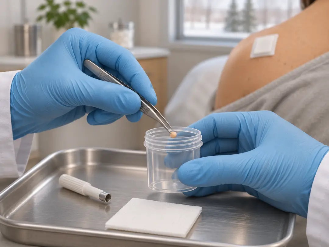
At a glance
- Visit
- About 15 to 30 minutes
- Healing
- About 1 to 2 weeks
- Anaesthetic
- Local anaesthetic
- OHIP
- OHIP-covered
Yes for suspicious moles. Cosmetic removal is not.
No referral needed.
A skin biopsy is how that worry gets a clear answer. A small piece of skin, or the whole mole, is removed and checked under a microscope by a pathologist.
The good news: most biopsied moles turn out to be harmless. And when something does need treatment, finding it early makes a real difference.
Assessment before treatment. Every mole and skin lesion is assessed before any treatment. The assessment combines Dr. Bastien's clinical experience and dermatology training with dermoscopy, digital imaging and AI-assisted analysis where appropriate.
Assessed first
Every mole is examined with dermoscopy before deciding whether to remove it
Quick and in clinic
Most biopsies take 15 to 30 minutes under local freezing
Lab-confirmed
Every removed mole of concern is sent for testing
Quick answers
Does it hurt?
Only the freezing needle stings briefly. Then you feel pressure.
How long does it take?
About 15 to 30 minutes
When are results back?
Usually 1 to 2 weeks
Healing time?
About 1 to 2 weeks. Longer on the lower legs.
Will it scar?
A small scar is normal and fades over months
Is it covered by OHIP?
Yes for suspicious moles. Cosmetic removal is not.
Do I need a referral?
No
When does a mole need a biopsy?
Most moles never need removing. A biopsy is advised when a mole looks unusual, has changed, or cannot be confidently judged on examination alone.
Warning signs: the ABCDEs of melanoma
- A
Asymmetry. One half does not match the other
- B
Border. Ragged, notched or blurred edges
- C
Colour. Several shades of brown, black, red, white or blue
- D
Diameter. Larger than about 6 mm, though melanomas can be smaller
- E
Evolving. Any change in size, shape, colour or height
Other signs worth checking
- A new mole after age 30, or one that looks different from your other moles (the "ugly duckling" sign)
- A mole that itches, bleeds, crusts or will not heal
- A dark streak under a nail
Where do cancerous moles appear? In men, melanoma is most common on the back. In women, it is most common on the legs. In darker skin, it more often appears on the palms, soles and under the nails.
Can a doctor tell if a mole is cancer just by looking?
An experienced eye and a dermoscope give a lot of information. Dermoscopy magnifies the mole and shows patterns invisible to the naked eye.
This helps separate moles that look reassuring from those that need testing. But only a biopsy examined in a lab can confirm whether a mole is cancer.
Most biopsied moles are benign. Biopsying a mole is about being certain, not about expecting the worst.
Mole removal and biopsy methods
The method depends on what the mole looks like, its size and where it is.
Shave biopsy or removal
How it is done: A small blade removes the mole level with the skin
Stitches? No
Often used for: Raised moles that look benign
Punch biopsy
How it is done: A round tool removes a small core of skin
Stitches? Usually 1 to 2
Often used for: Sampling part of a larger spot
Excisional biopsy
How it is done: The whole mole is cut out with a small margin
Stitches? Yes
Often used for: Moles that could be melanoma
Suspicious moles are usually removed whole. When melanoma is a concern, removing the entire mole gives the lab the most accurate picture.
Can moles be removed with a laser? A mole that may need testing should not be lasered, because laser destroys the tissue and nothing is left to examine.
What happens during the procedure
- 1
Exam
The mole is examined with dermoscopy and photographed
- 2
Consent
You review the method, scarring and what results mean
- 3
Freezing
Local anaesthetic is injected. It stings for a few seconds.
- 4
Removal
The mole is shaved, punched or excised. You feel pressure only.
- 5
Closure
Stitches if needed, then ointment and a bandage
Most procedures take 15 to 30 minutes, and you can return to work or daily activities the same day.
Not sure this is the right treatment?
Every spot is assessed before anything is treated. If another option suits you better, Dr. Bastien will tell you.
Healing and aftercare
- 1
First 24 to 48 hours
Keep the bandage on and dry. Mild soreness is normal.
- 2
Days 2 to 14
Wash gently once a day, apply petroleum jelly and re-cover
- 3
Stitches out
About 5 to 7 days on the face, 10 to 14 days on the body
- 4
1 to 2 weeks
Most wounds have closed. Lower legs can take longer.
- 5
Months 1 to 12
The scar flattens and fades
Avoid swimming, baths and hot tubs, and stretching the area, until the wound has closed or stitches are out.
Call the clinic if bleeding does not stop after 20 minutes of firm pressure, or you notice spreading redness, pus, worsening pain or fever.
To help the scar fade: keep the wound moist while healing, use silicone gel once closed, and protect it from the sun for a year.
Understanding your biopsy results
Results usually take about 1 to 2 weeks. Dr. Bastien will contact you or review them at a follow-up visit.
Benign mole
What it means: Harmless. Usual next step: No further treatment.
Atypical (dysplastic) mole
What it means: Unusual cells, not cancer. Usual next step: Sometimes a small re-excision for a clear margin.
Basal or squamous cell carcinoma
What it means: A common, treatable skin cancer. Usual next step: Excision or another treatment, often in clinic.
Melanoma
What it means: A skin cancer that can spread. Usual next step: Prompt wider excision and further care, with referral if needed.
If a biopsy shows melanoma, the next step is usually a wider excision around the scar to make sure it is fully removed. Thicker melanomas may need more tests. Early, thin melanomas have the best outcomes, which is why prompt testing matters.
Why do moles sometimes grow back?
A mole that is shaved off can leave pigment cells deeper in the skin. These can slowly bring back some colour at the same spot.
This is usually harmless, but any regrowth should be shown to your doctor. Full excision gives the lowest chance of a mole returning.
Home remedies and creams that claim to "kill" moles are not safe. They can burn the skin and destroy a mole before it can be tested.
Related treatments
- Mole MappingPhotos of your whole skin and close-ups of single moles, compared at each visit so Dr. Bastien can see what is new or has changed.
- Skin Cancer AssessmentA doctor examines the spot that worries you, and you leave with a plan: watch it, test it or treat it.
- Skin Cancer SurgeryDr. Bastien removes the skin cancer in the clinic under local freezing, then closes the wound with stitches or a skin flap.
Frequently asked questions
Is mole removal the same as a skin biopsy?
Often, yes. When a whole mole is removed and sent to the lab, the removal is also the biopsy. A punch biopsy only samples part of a spot.
Does a mole biopsy hurt?
The freezing needle stings for a few seconds. After that, you should feel only pressure. Soreness afterwards is usually mild.
Is a skin biopsy only done for cancer?
No. Biopsies also help diagnose rashes, infections and other skin conditions. For moles, they confirm whether a spot is harmless.
What percentage of mole biopsies are cancerous?
Most biopsied moles are benign. Only a small share turn out to be melanoma, but testing is the only way to be sure.
Will a biopsy leave a scar?
Yes, a small scar is normal. Shave removals leave a flat, pale mark. Excisions leave a fine line that fades over months.
How quickly can melanoma progress?
It varies. Some melanomas grow slowly over years, while others change within weeks to months. Any changing mole should be checked promptly.
Is mole removal safe?
Yes, when done by a trained clinician with proper testing. Risks such as infection or bleeding are uncommon.
Do I need a referral?
No. You can book directly at 1692 Murphy Rd, Sarnia, by calling 1-866-550-MOLE (6653).
Book a consultation
You don’t need a referral. Dr. Bastien examines the area first and tells you whether you need treatment.
1692 Murphy Rd, Sarnia
Monday to Friday, 8:30 am to 4:30 pm
Free parking on site