Curettage and Electrodesiccation in Sarnia, Ontario
Curettage and electrodesiccation (often shortened to ED&C) is an in-clinic procedure.
At a glance
- Healing
- About 3 to 6 weeks
- Anaesthetic
- Local anaesthetic
- OHIP
- OHIP-covered
Usually, when done for a medical reason
No referral needed.
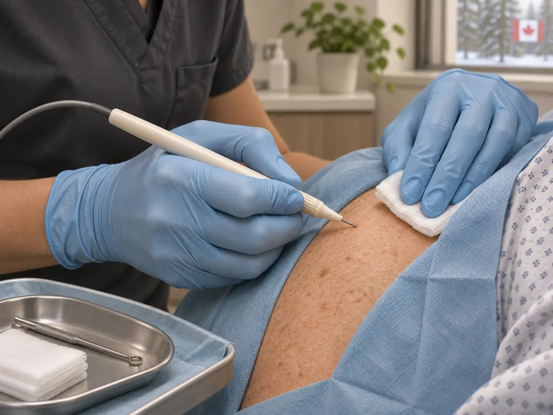
Curettage and electrodesiccation (often shortened to ED&C) is an in-clinic procedure. The doctor scrapes away a lesion with a small ring-shaped tool, then uses heat on the base to treat the remaining cells and stop bleeding. It is used for selected small, low-risk skin cancers and for some other growths.
Assessment first
The lesion is examined, and a biopsy confirms the diagnosis before a skin cancer is treated
Local anesthetic
The area is numbed, and you stay awake
Quick
A typical lesion takes about 10 to 20 minutes
No stitches
The wound heals from the base over several weeks
For selected lesions only
Not used for melanoma, or for deeper or higher-risk cancers
Quick answers
What does it treat?
Small, low-risk superficial basal cell carcinoma, SCC in situ, and some benign growths and thick sun-damage patches
Does it hurt?
Injecting the anesthetic stings. The procedure itself is not painful.
How long does healing take?
About 3 to 6 weeks, and longer on the lower legs
Will it leave a scar?
Yes. It is usually round, flat and paler than the surrounding skin.
Is the tissue tested?
A biopsy is taken first. The scraped tissue gives no margin check.
Can it come back?
Yes. Follow-up checks are advised.
Is it covered by OHIP?
Usually, when done for a medical reason
What is curettage and electrodesiccation, and how does it work?
A curette is a small, sharp, ring-shaped instrument. Soft tumour tissue is more crumbly than firm, healthy skin, so the doctor can feel when the abnormal tissue has been scraped away. This is why the technique relies on the doctor's skill and experience.
Electrodesiccation then uses a small electric current to dry out and destroy any cells that are left on the base and edges of the wound, and to seal small blood vessels. The scraping and the heat are often repeated two or three times in the same visit.
Curettage alone
Sometimes the scraping is done without the heat step. For some small, low-risk lesions, curettage alone has been reported to have a similar success rate to curettage with electrodesiccation, with less scarring. The doctor decides which method suits the lesion.
What it treats, and where it is not used
All skin lesions are assessed by a doctor before treatment. The assessment combines clinical experience, dermoscopy and digital imaging, with AI-assisted analysis where appropriate. A biopsy confirms the diagnosis before a skin cancer is scraped.
Small, superficial or nodular basal cell carcinoma with low-risk features
A mole, or any spot that could be a melanoma.
SCC in situ (Bowen’s disease)
Morpheaform or other high-risk basal cell carcinoma.
Thin, low-risk SCC on the trunk and limbs
Deep, large or returned skin cancers.
Thick or stubborn actinic keratoses
Cancers on the lip, eyelid, nose or ear that may need Mohs surgery.
Seborrheic keratoses, warts, skin tags
Skin where tumour cells can follow hair follicles deep, such as the scalp, beard area and underarms.
Some pyogenic granulomas and other small benign growths
A tumour that reaches into fat when scraped. The plan then switches to excision.
Why a biopsy comes first
Scraping gives no piece of tissue with its edges intact for a lab to check. A biopsy before treatment tells the doctor what the lesion is and whether it is low-risk enough for this method.
Tell the clinic if you have
- A pacemaker or a defibrillator, because the heat tool may need to be changed or avoided
- A bleeding problem, or you take blood thinners
- A history of keloid or raised scars
- Allergies, including to local anesthetic
The visit and the procedure, step by step
- 1
Check
The lesion is re-examined and the plan is confirmed. Consent is reviewed and signed.
- 2
Numbing
Local anesthetic is injected. It stings for a few seconds, then the area goes numb.
- 3
Curettage
The curette scrapes the lesion until firm, healthy-feeling skin is reached. You feel pressure and scraping, not pain.
- 4
Electrodesiccation
A fine electrical tip is touched to the base and edges. It stops bleeding and treats remaining cells. You may smell a slight odour.
- 5
Repeat
Scraping and heat are repeated once or twice, if needed
- 6
Dressing
Ointment and a dressing are applied, and written aftercare is given
The procedure takes about 10 to 20 minutes for one lesion, and the visit takes longer with the assessment and instructions. Most people drive home and return to normal activities the same day, unless the lesion is near the eye.
Not sure this is the right treatment?
Every spot is assessed before anything is treated. If another option suits you better, Dr. Bastien will tell you.
Healing and wound care
The wound is left open and heals from the base up. It does not need stitches.
- 1
Day 0 to 2
Mild soreness. Some oozing or blood-tinged fluid.
What to do: Keep the dressing on and dry as instructed
- 2
Day 3 to 14
A scab or crust forms and the wound may weep
What to do: Wash gently, pat dry, apply ointment and a fresh dressing
- 3
Weeks 2 to 6
The scab falls off and pink new skin shows
What to do: Do not pick the scab. Protect the area from the sun.
- 4
Months 3 to 12
The scar flattens and the colour changes
What to do: Use sunscreen on the scar
Aftercare
- Acetaminophen is usually enough for soreness
- Keep the wound moist and covered with the ointment advised, as a dry scab can slow healing
- Do not soak the area in a bath, pool or lake while the wound is open
- Avoid heavy exercise if the wound is on the trunk or limbs and pulls open
Call the clinic if
- Redness spreads or the pain gets worse after the second day
- Pus drains from the wound, or you have a fever
- Bleeding does not stop with firm pressure
- The wound has not healed after 6 weeks
Scarring, colour change, risks and follow-up
Round scar
Typically flat, round and pale. It has been compared with a small burn mark.
Lighter skin
Common. It is more noticeable in darker skin.
Raised or thick scar
Possible, more likely on the chest, shoulders and upper back.
Bleeding
Usually small and stopped by firm pressure.
Infection
Uncommon.
Slow healing
More likely on the lower legs, and in smokers.
Recurrence
The lesion can come back, and the chance depends on the type, size and place.
Follow-up
A recheck of the site is booked after healing, and regular skin checks are advised. Because the method leaves no piece of tissue with its edges for the lab, any area that looks unusual during healing, or that returns, is re-examined and may be biopsied or treated by another method.
People who have had one skin cancer are more likely to develop another. A whole-body check with photos can help track new or changing spots.
Compared with surgery, cryotherapy, PDT and Mohs surgery
Reported five-year cure rates for small, first-time basal cell carcinomas are above 95% for curettage and electrodesiccation and for excision. Rates vary between studies and depend on the type, size and place.
Surgical excision
Cuts out the lesion with a margin and sends it to the lab. A line scar and stitches. Suits more lesion types and sizes.
Cryotherapy
Freezes the lesion. No anesthetic is usually needed. Suited to thin lesions. Blisters and pale marks.
Photodynamic therapy
Cream and a lamp. Suits flat superficial lesions and large areas. Little scarring.
Mohs surgery
Removes the cancer in layers with a lab check of the edges during the visit. For cancers in high-risk places, and large or returned ones. By referral.
Curettage and electrodesiccation
Quick and done in one visit. Round pale scar. No margin check.
The right method is chosen after the biopsy and examination, using the type, size, depth and place, and your own preferences.
Cost and OHIP
When the procedure treats a biopsy-confirmed skin cancer or another medical problem, it is generally billed to OHIP. Cosmetic removal of harmless growths is usually not covered.
Related treatments
- Basal Cell Carcinoma TreatmentDiagnosis and in-clinic treatment of basal cell carcinoma, the most common skin cancer, by surgery, scraping, freezing or light therapy.
- Squamous Cell Carcinoma TreatmentDiagnosis and treatment of squamous cell carcinoma and SCC in situ (Bowen’s disease), most often by surgical removal.
- Skin Cancer SurgeryDr. Bastien removes the skin cancer in the clinic under local freezing, then closes the wound with stitches or a skin flap.
Frequently asked questions
Is curettage and electrodesiccation painful?
The local anesthetic injection stings for a few seconds. After that, you feel pressure and scraping, but not pain. Soreness after the numbness wears off is usually mild and is managed with acetaminophen.
How long does the wound take to heal?
Usually about 3 to 6 weeks, and longer on the lower legs. The wound scabs over and heals from the base. The scar keeps changing for months.
Will it leave a scar?
Yes. The scar is usually round, flat and paler than the skin around it. Raised scars can occur, especially on the chest and upper back.
Why do I need a biopsy first?
The procedure leaves no tissue with its edges for the lab. The biopsy tells the doctor whether the lesion is a skin cancer, what type it is and whether this method suits it.
Can it treat any skin cancer?
No. It is used for small, low-risk lesions, such as some superficial basal cell carcinomas and SCC in situ. Melanoma, high-risk cancers, and cancers in some places need surgery or another treatment.
What is the difference between curettage and electrodesiccation?
Curettage is the scraping with a small tool. Electrodesiccation is the heat step that dries the base and stops bleeding. They are often used together, and sometimes curettage is used alone.
Can the lesion come back?
Yes. The chance depends on the type, size and place. Follow-up checks are advised, and any area that changes or returns should be seen promptly.
Can I go back to work and exercise?
Most people go back to work the same day or the next. Avoid heavy exercise or swimming while the wound is open, particularly if it is on a place that stretches, such as the back or shoulders.
Book a consultation
You don’t need a referral. Dr. Bastien examines the area first and tells you whether you need treatment.
1692 Murphy Rd, Sarnia
Monday to Friday, 8:30 am to 4:30 pm
Free parking on site