Basal Cell Carcinoma Treatment in Sarnia, Ontario
Basal cell carcinoma (BCC) is the most common skin cancer. It grows slowly, usually stays in one place and rarely spreads. It still needs treatment, because it keeps growing and can damage the skin around it.
At a glance
- OHIP
- OHIP-covered
Usually, when it is medically necessary
No referral needed.
Common
BCC makes up about 75 to 80% of non-melanoma skin cancers in Canada
Often on sun-exposed skin
Face, scalp, ears, neck, shoulders and back
Confirmed by biopsy
The type is checked before treatment is chosen
Several treatments work
Surgery, scraping and heat, freezing, light treatment, creams or radiation
Treated in clinic when suitable
Some cases are referred, for example certain cancers near the eye, nose or ear
Quick answers
Can BCC be treated successfully?
Most small BCCs found early are treated successfully, and the doctor explains the chance for your type and place
Does it spread?
Spread to other organs is rare. Local growth is the main concern.
Do I need a biopsy?
Usually yes, to confirm the type before treatment
What is the most common treatment?
Surgical excision, or scraping with heat (curettage and electrodesiccation) for small, low-risk lesions
Will it leave a scar?
Yes, every method leaves some mark. Its shape depends on the method.
Can it come back?
Yes. Follow-up checks are advised.
Is it covered by OHIP?
Usually, when it is medically necessary
What is basal cell carcinoma?
BCC starts in the basal cells, which sit in the deepest layer of the top skin layer (the epidermis). It is a non-melanoma skin cancer. It usually grows slowly over months or years.
The main cause is ultraviolet (UV) light from the sun and tanning beds. UV damages the DNA of skin cells over time.
Who is more likely to get BCC
- People with fair skin, light eyes or hair that burns easily
- People with years of sun exposure, including outdoor work
- People with a history of sunburns, tanning-bed use or earlier skin cancer
- Older adults, although it is now seen in younger people too
- People who have had radiation treatment to the skin
- People with a weakened immune system
Anyone can develop skin cancer, whatever their skin colour. A spot that does not heal should be checked in any skin colour.
Non-melanoma skin cancers in Ontario rose by about 30% between 2003 and 2017, based on a study in the Canadian Medical Association Journal.
What can basal cell carcinoma look like?
BCC can look quite different from one person to another. Only an examination can tell it apart from harmless spots, so this list is a guide to when to book a check, not a way to diagnose.
Nodular
A pearly or pink bump, often with tiny visible blood vessels. It may have a rolled edge and a central dip.
Superficial
A flat, pink or red patch that may be scaly. It can look like eczema and does not clear.
Pigmented
A brown, blue or black bump that can be mistaken for a mole.
Morpheaform (infiltrative)
A flat, firm, shiny area that looks like a scar and has no clear edge.
Ulcerated
A sore that bleeds, crusts and does not heal in 4 weeks.
Book a check if you notice
- A sore that bleeds, scabs and does not heal
- A shiny bump that slowly grows
- A red, scaly patch that does not respond to moisturizer or creams
- A scar-like area that you do not remember an injury for
- A mole-like spot that is new or changing
BCC is often painless, so the lack of pain does not mean a spot is harmless. See a clinician rather than waiting.
How basal cell carcinoma is assessed and diagnosed
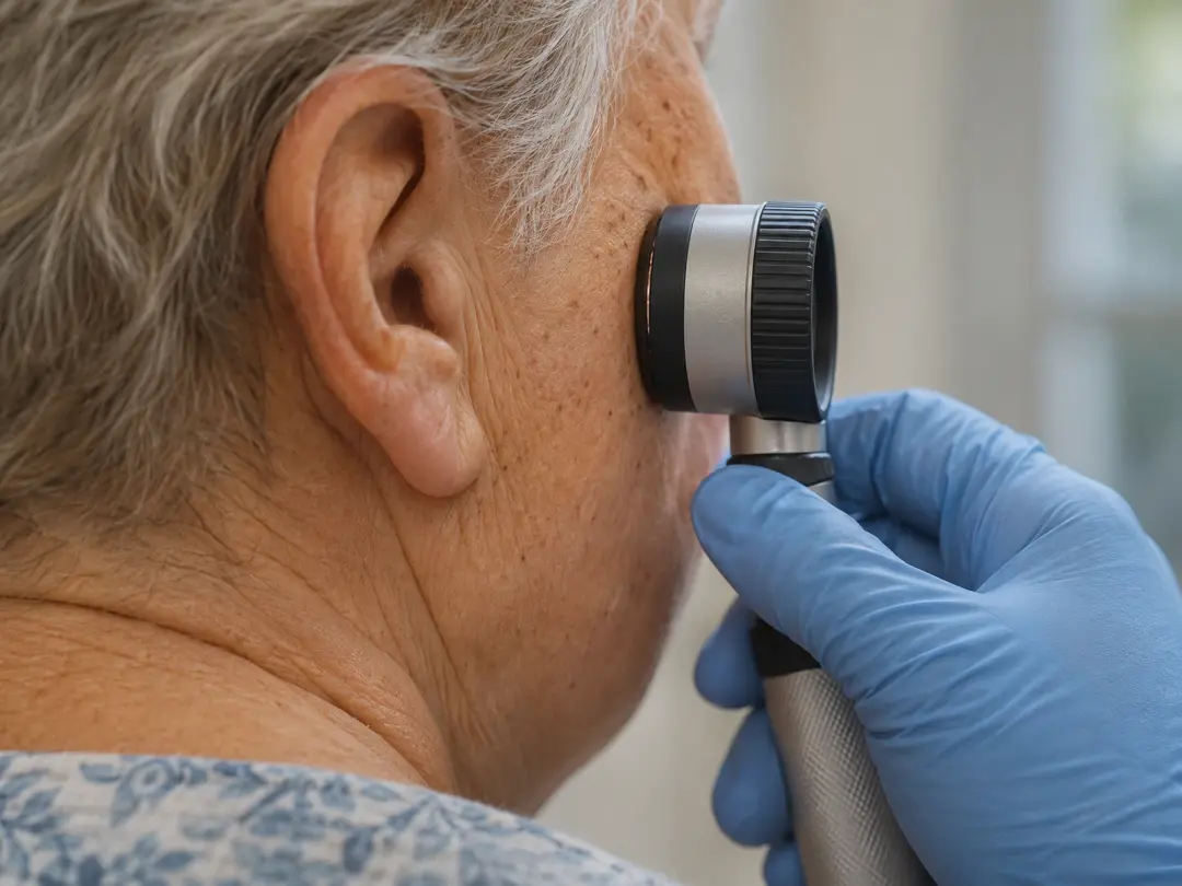
All skin lesions are assessed by a doctor before treatment. The assessment combines clinical experience, dermoscopy and digital imaging. AI-assisted analysis is used where appropriate, alongside the doctor's own judgement.
- 1
History
When the spot appeared, whether it bleeds or grows, sun exposure and any earlier skin cancer
- 2
Examination
The spot and the rest of the skin are checked, because there may be more than one
- 3
Dermoscopy
A magnifying, lit device shows patterns that are typical of BCC, such as fine branching vessels
- 4
Biopsy
A small sample is taken under local anesthetic and sent to a lab. It confirms the diagnosis and the subtype.
- 5
Plan
The result is explained and treatment options are discussed
The biopsy report gives the subtype. Superficial and nodular BCCs are often considered lower risk. Morpheaform and other infiltrating types are higher risk, because their edges are hard to see and they can reach deeper than they look.
Size, place on the body, and whether the spot has been treated before also affect the choice of treatment.
Not sure this is the right treatment?
Every spot is assessed before anything is treated. If another option suits you better, Dr. Bastien will tell you.
Treatment options for basal cell carcinoma
Many BCCs can be treated in an outpatient clinic under local anesthetic. Reported five-year success rates for small, first-time BCCs are above 90% for surgery, scraping with heat, freezing and radiation, and lower for creams. The figures vary between studies and depend on the type, size and place.
Surgical excision
Usually considered for: Most BCC types and sizes, including those that need the edges checked
Points to know: A line scar. Stitches. The lab checks the margins.
Curettage and electrodesiccation
Usually considered for: Small, low-risk, superficial or nodular BCC on the trunk and limbs
Points to know: Quick. Round pale scar. No margin check.
Cryotherapy
Usually considered for: Selected thin, small, low-risk BCC
Points to know: Blister and a pale mark are common. No margin check.
Photodynamic therapy (PDT)
Usually considered for: Selected superficial BCC
Points to know: A confirmed diagnosis is needed. Red, sore skin for several days.
Mohs surgery
Usually considered for: BCC near the eyes, nose, lips or ears, and large, aggressive or returned BCC
Points to know: Done by a Mohs surgeon. Needs a referral.
Creams (imiquimod, fluorouracil)
Usually considered for: Some superficial BCC
Points to know: Used for weeks. Skin gets red and crusted. Lower success rates than surgery.
Radiation therapy
Usually considered for: Older patients or places where surgery is difficult
Points to know: Several visits over a few weeks. Done by a radiation team.
How the choice is made
- The subtype and risk level on the biopsy report
- The size and depth of the tumour
- The place on the body and how it will heal there
- Whether it has been treated before
- Your health, medicines and preferences
You are involved in the choice. Questions about scarring, healing time and follow-up are part of the discussion.
How each option is done at the clinic
Surgical excision. After local anesthetic, the doctor cuts out the BCC with a margin of normal-looking skin. The wound is closed with stitches, or with a local skin flap if it is too large to close in a line. The tissue goes to the lab. See the page on skin cancer surgery for the full steps.
Curettage and electrodesiccation. After local anesthetic, a small ring-shaped tool scrapes away the soft tumour. Heat is then used to treat the base and stop bleeding. This is repeated once or twice in the same visit. The wound heals on its own over several weeks. Curettage is not used for BCC that reaches into fat, for morpheaform BCC, or for BCC that has come back.
Cryotherapy. Liquid nitrogen is applied to freeze the lesion and a small ring around it. The freeze lasts a few seconds to a minute, and may be repeated. A blister forms and then crusts. Cryotherapy is limited to selected lesions after assessment and, when needed, a biopsy.
Photodynamic therapy. A light-sensitising cream is applied, left to absorb, and then the area is lit with a special lamp. It is used for selected superficial BCC and is described on its own page.
When the clinic refers
- BCC near the eyelid, nose, lip or ear
- Large, deep, aggressive or returned BCC
- Cases where Mohs surgery, a graft or radiation is the more suitable option
A referral is made after a biopsy confirms the diagnosis, and you are told why.
Recovery, scarring and aftercare
Healing depends on the method used and the place treated.
Excision with stitches
Healing: Stitches out in about 5 to 14 days. The scar matures over 9 to 12 months.
Scar: A line, often longer than the BCC
Curettage and electrodesiccation
Healing: An open wound that scabs and heals over about 3 to 6 weeks
Scar: A round, pale, flat scar
Cryotherapy
Healing: A blister, then a crust that falls off in 1 to 3 weeks. Full healing can take longer.
Scar: A pale mark. It may be slightly sunken.
PDT
Healing: Redness, crusting and soreness for about 1 week
Scar: Usually little scarring
Creams
Healing: Redness and crusting during treatment
Scar: Usually little scarring
Aftercare in general
- Keep the dressing clean and dry for the first 24 to 48 hours, or as instructed
- Wash gently with water, pat dry and apply the ointment you are given
- Do not pick scabs
- Do not soak the area until the wound is closed or the stitches are out
- Protect the site from the sun for at least a year, as new scars burn easily
- Call the clinic for spreading redness, pus, fever or bleeding that does not stop with firm pressure
Pale or darker marks can occur with any method. They are more likely with freezing and heat.
Recurrence, follow-up and prevention
BCC can come back in the same place, and people who have had one BCC are more likely to develop another elsewhere. The chance depends on the subtype, size, place and the treatment used.
Follow-up
- A recheck of the treated site, usually within a few weeks to months
- Regular full-skin checks. The interval is set by your doctor, and many people are checked every 6 to 12 months at first.
- Whole-body photos and mole mapping can help track new or changing spots
Can BCC spread?
Spread to other parts of the body is rare. The main concern is local growth, which can damage nearby skin, and in some places cartilage or bone, if a BCC is left for a long time.
Lowering your risk of another one
- Use a broad-spectrum sunscreen of SPF 30 or higher on exposed skin, and reapply it
- Wear a hat, sunglasses and long sleeves in strong sun. Sarnia’s lakeshore reflects sunlight, so shade and clothing matter on the water and the beach too.
- Avoid tanning beds
- Check your own skin monthly, including the scalp, ears, back and legs, with a mirror or a helper
- Book a check for any new or changing spot
Cost and OHIP
Treatment of a biopsy-confirmed basal cell carcinoma is usually medically necessary and is generally billed to OHIP. Coverage can differ for some items, so the clinic confirms what applies before treatment.
Who treats basal cell carcinoma in Sarnia?
Dr. David Bastien, MD, MBA, DipPDerm (Cardiff), assesses and treats skin cancers at Dr. Bastien Mole Clinic on Murphy Road in Sarnia. He holds a CPSO-approved change of scope to Skin Disorders and Ontario Medical Association Focused Practice recognition in Dermatology.
The clinic offers assessment with dermoscopy and digital imaging, biopsy, surgical excision, curettage and electrodesiccation, cryotherapy and photodynamic therapy for suitable lesions.
Referral to another team
Some BCCs are better treated by a Mohs surgeon, a plastic surgeon or a radiation team. When that is the case, the clinic explains why, sends the referral and shares the biopsy report.
See the About Dr. Bastien page for his training.
Related treatments
- Skin Cancer SurgeryDr. Bastien removes the skin cancer in the clinic under local freezing, then closes the wound with stitches or a skin flap.
- Curettage and ElectrodesiccationScraping and heat for small, low-risk skin cancers and some harmless growths. The wound heals without stitches.
- CryotherapyFreezing with liquid nitrogen for sun-damage patches, seborrheic keratoses, skin tags and selected early skin cancers.
- Photodynamic Therapy (PDT)A light-activated cream that treats many sun-damage patches on the face or scalp in one session.
Frequently asked questions
Is basal cell carcinoma serious?
BCC is the least likely skin cancer to spread, but it is still a cancer. It keeps growing and can damage the skin, and sometimes the tissue beneath, if it is not treated. Early treatment usually means a smaller treatment and a smaller scar.
Can I just watch a basal cell carcinoma?
BCC does not go away on its own. A spot that is biopsy-confirmed as BCC is usually treated. In a few cases, such as older age or ill health, a doctor may discuss other plans with you.
What is the difference between basal cell and squamous cell carcinoma?
They start in different skin cells. BCC grows slowly and rarely spreads. Squamous cell carcinoma is less common, grows faster in some cases and is more likely to spread, so it is assessed with extra care.
Do I need a biopsy before treatment?
Usually yes. A biopsy confirms the diagnosis and shows the subtype, which helps choose the method. It is done under local anesthetic.
Which treatment leaves the smallest scar?
Every method leaves a mark. Scars from surgery are lines, and scars from scraping or freezing are round and pale. The right choice depends on the type, size and place, so it is decided at the assessment.
How long does the wound take to heal?
Stitches come out in about 5 to 14 days. A wound left open after scraping heals over about 3 to 6 weeks. The scar keeps changing for 9 to 12 months.
Can a basal cell carcinoma come back after treatment?
Yes, it can return in the same place, and new ones can appear elsewhere. Follow-up skin checks are advised, and any new or changing spot should be seen promptly.
Do I need a referral to book?
No. You can book an assessment directly. If a Mohs surgeon or another team is the better option, the clinic arranges the referral.
Book a consultation
You don’t need a referral. Dr. Bastien examines the area first and tells you whether you need treatment.
1692 Murphy Rd, Sarnia
Monday to Friday, 8:30 am to 4:30 pm
Free parking on site